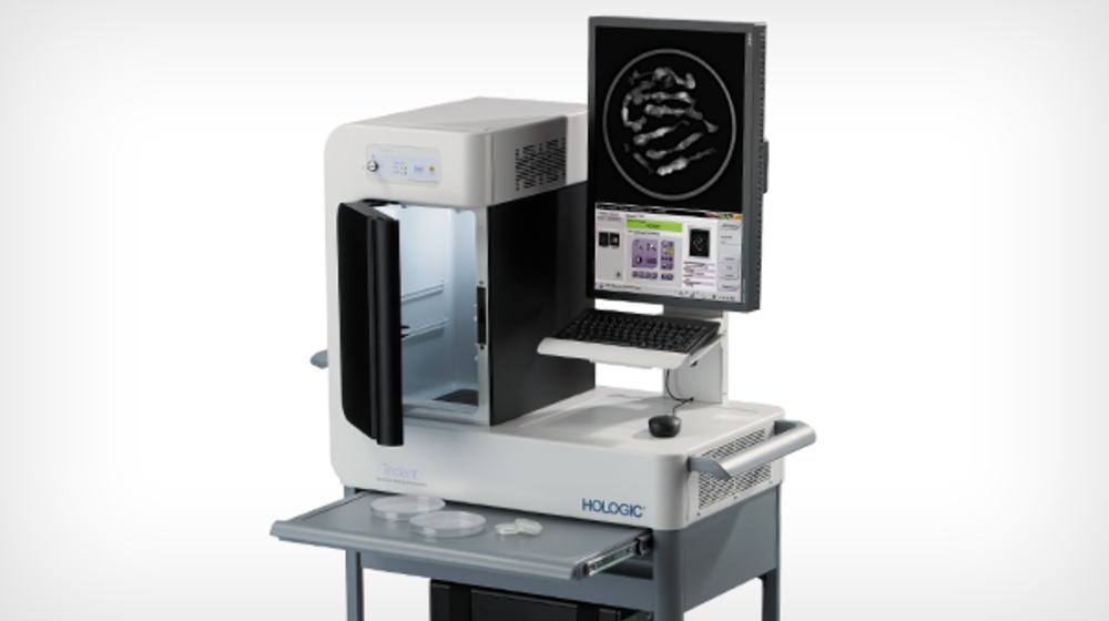
Trident™ Specimen Radiography System
Quality images, quickly delivered
This system provides exceptional breast tissue imaging, with a micro-focused tube, amorphous selenium direct digital detector and proprietary specimen image processing.
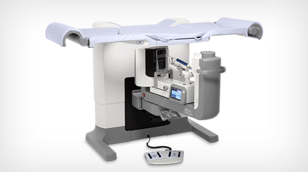
Stereotactic
Features
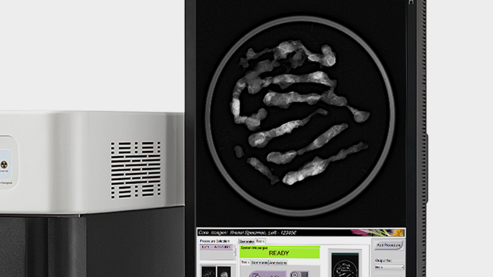
Exceptional image quality
Algorithms optimized for breast sample radiography provide high-resolution images via a 12cm x 14cm imaging area, utilizing our proprietary selenium, direct-conversion technology.
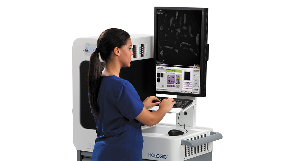
Simplified operation
Intuitive interface and software, ideal for non-technical users, with automatic exposure control and extensive tool set.
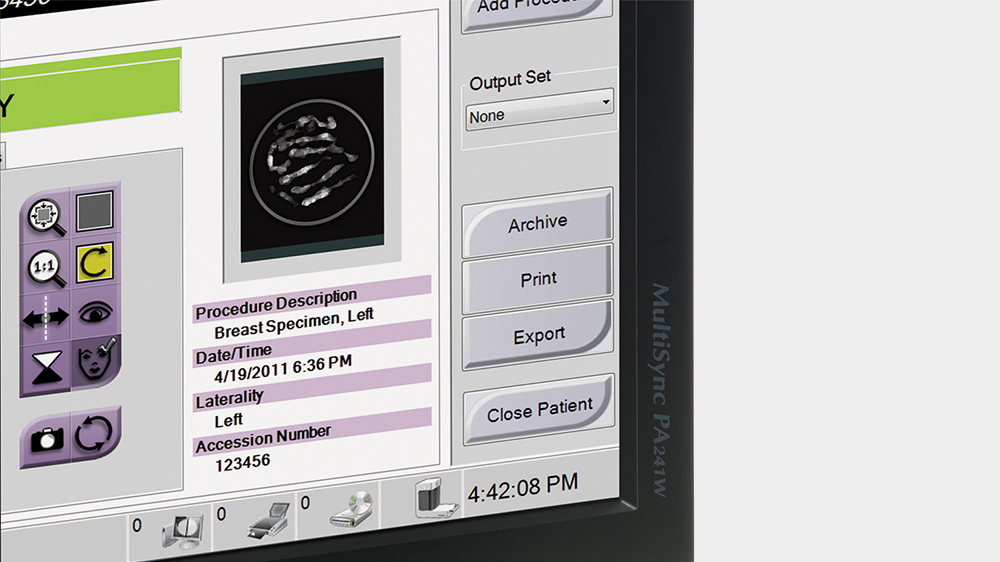
Robust connectivity
One-button send to SecurView DX, PACS, printer or external media devices. Annotations, measurements and enhancements saved in new DICOM file.
Your Complete Biopsy Solution
Choose Your Method:
Stereotactic
Ultrasound
MRI