Trident Specimen Radiography System

Quality images, quickly delivered

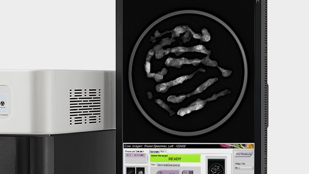
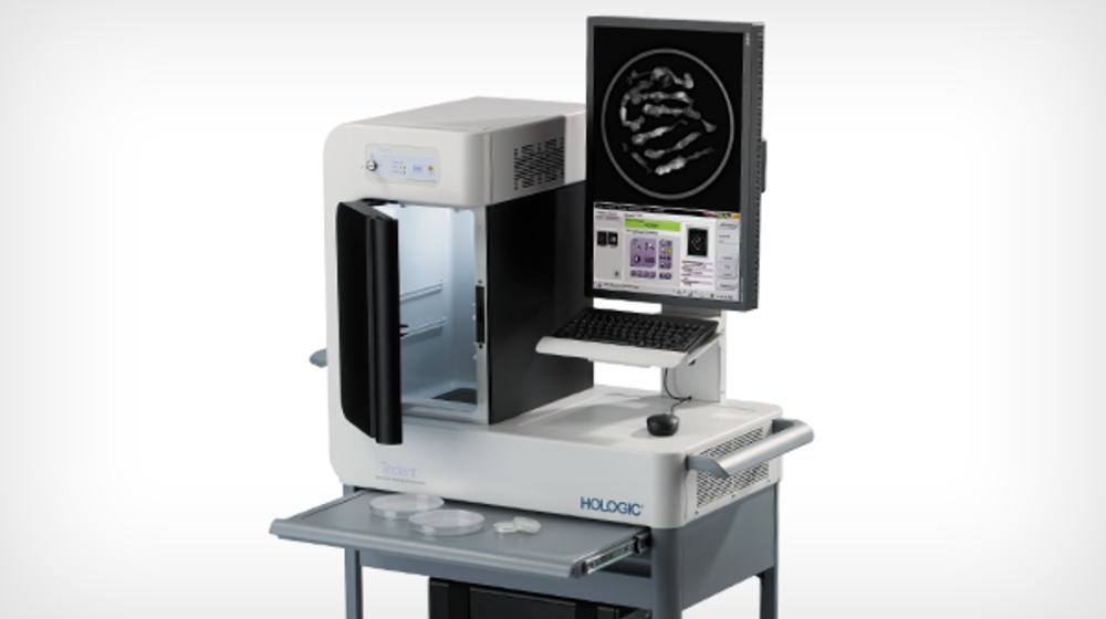
This system provides exceptional breast tissue imaging, with a micro-focused tube, amorphous selenium direct digital detector and proprietary specimen image processing.

Stereotactic

Features

Exceptional Image Quality

Exceptional image quality

Algorithms optimized for breast sample radiography provide high-resolution images via a 12cm x 14cm imaging area, utilizing our proprietary selenium, direct-conversion technology.
Simplified operation

Simplified operation

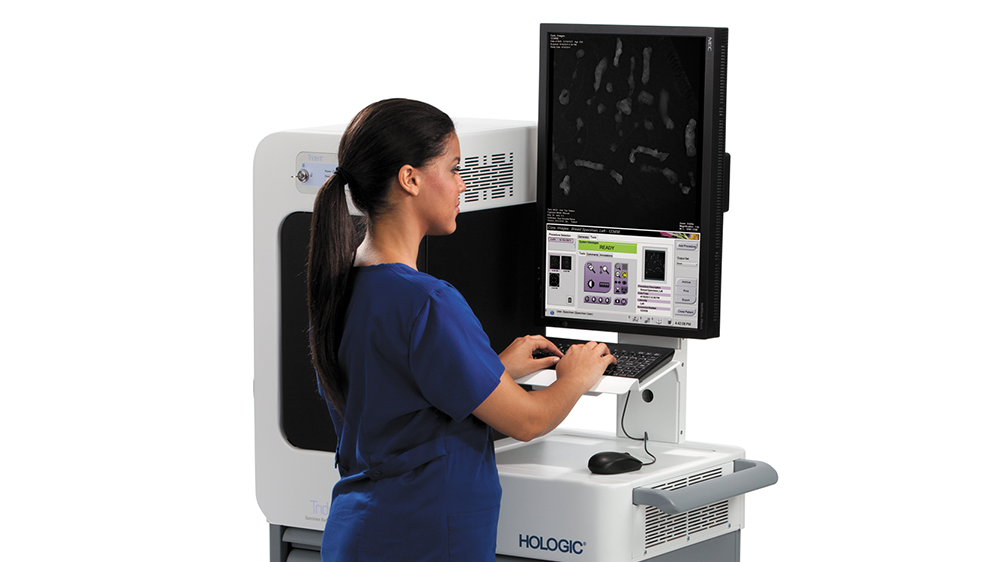
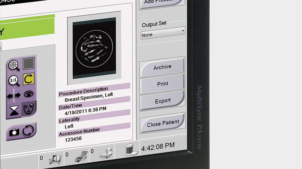
Intuitive interface and software, ideal for non-technical users, with automatic exposure control and extensive tool set.
Robust connectivity

Robust connectivity

One-button send to SecurView DX, PACS, printer or external media devices. Annotations, measurements and enhancements saved in new DICOM file.
Tech Specs
Tech Specs  
Electrical Requirements  
Input Line Voltage 100-240 VAC
Peak Input Current 4 Amps
Frequency 50/60 Hz
Physical  
Construction Shielded cabinet on moveable cart
Dimensions 68.3” H , 37.5” W, 26.5” D
Weight 275 pounds
Detector Technology TFT-based direct capture technology
X-ray Absorption Material Amorphous selenium
Dimensions 68.3” H , 37.5” W, 26.5” D
Active Imaging Area Size 12 cm x 14 cm
Pixel Size 70 μm pixels
Limiting Spatial Resolution 7.1 lp/mm
Output Image 14-bit image data
Acquisition Workstation  
CPU Multi-core Intel
Memory 4 GB RAM
Hard Disk 500 GB
Storage Media DVD +/- R/W
Display Monitor 2.3 MP color LCD display
Operating System Windows 7 Professional
Network Connection 10, 100, 1000-Base-T; optional wireless
DICOM Services Worklist, print, storage
User Interface Keyboard, mouse
Image Display  
Image Processing Optimized display of core and surgical specimens, preset and manual window/level, invert
Visualization Enhancement Five user-selectable levels of image sharpening
Magnification Magnifying glass, continuous image zoom
Annotations Ellipse, polyline free-form, arrow, ruler, text box, comments
Multi-up 1-, 2- and 4-up
X-ray Source  
Energy Range 20-35 kV
Anode Type Tungsten
Tube Current 1 mA
Exposure Up to 10 mAs
Focal Spot Size 50 μm
Filtration 125 μm beryllium
Exposure Modes  
Manual User selects kV and mAs
Auto System determines optimum kV and mAs
System Performance  
Time to Preview < 20 seconds
Cycle Time 30 seconds
Cabinet  
Safety Features Door interlock, key switch, fully shielded
Indicators Power, door closed, ready, x-ray on
Magnification Specimen tray positions at 1.5x and 2.0x, auto-sensed
Other Lighted cabinet, easily cleaned
Accessories  
  Specimen tray
  User manual, quick start guide, service and cleaning
Optional Equipment  
  Uninterruptible Power Supply (UPS)

Your Complete Biopsy Solution

Choose Your Method: Stereotactic Ultrasound MRI

Looking for more information about the Trident
specimen radiography system?